

Advanced Imaging
Eye Associates NW has the latest imaging technology in ophthalmology. We can image the cornea at the front of the eye, to the retina at the back of the eye.
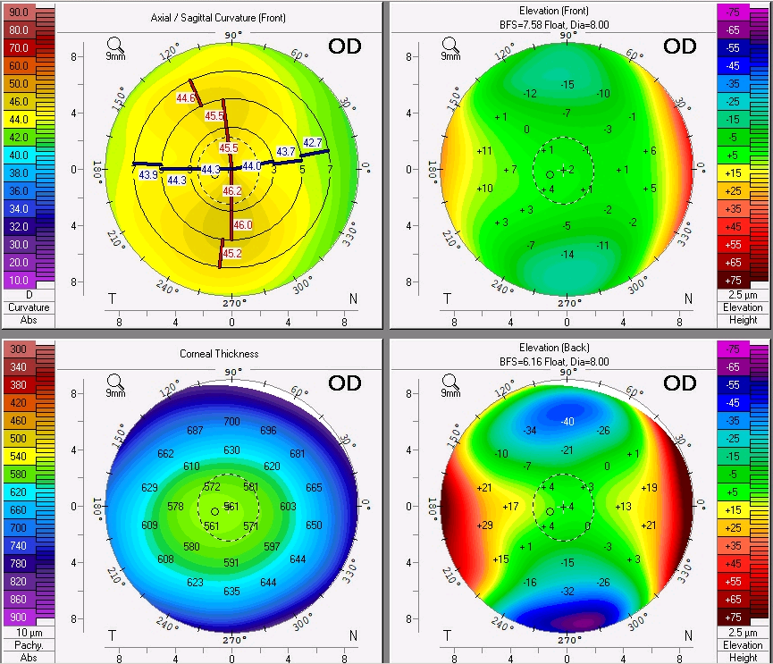
Corneal Topography / Pentacam
Corneal Topography and the Pentacam can image the front surface of the cornea to assess and follow corneal disease as well as the fitness for advanced intraocular lenses combined with cataract surgery. This is important for disorders such as Keratoconus and astigmatism management. The Pentacam can also asses the posterior surface of the cornea to prepare for laser refractive surgery. Advanced contact lens fitting is also assisted by these technologies.
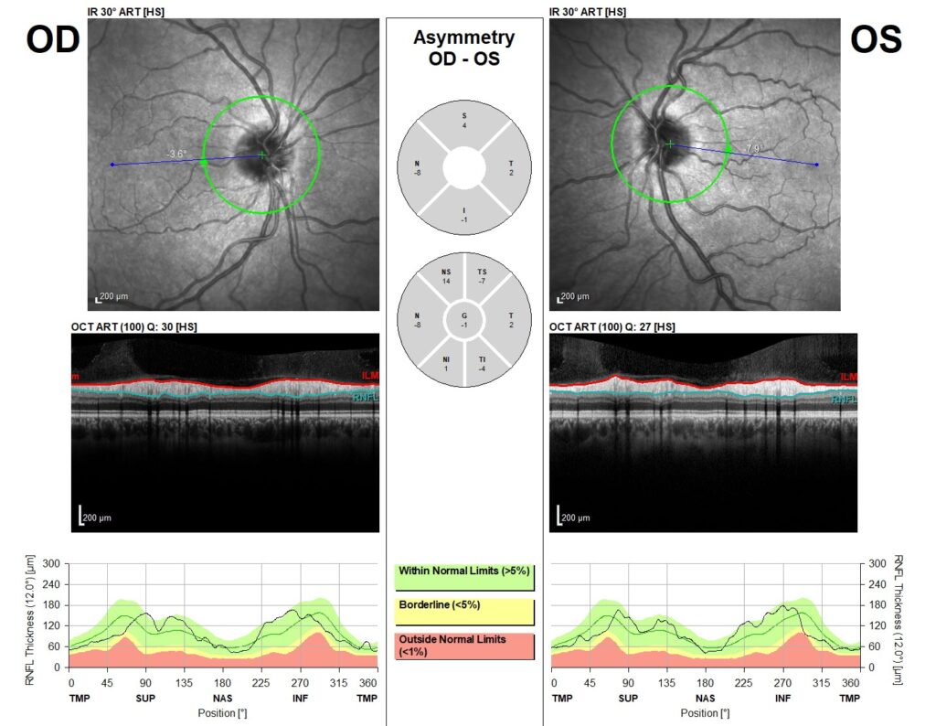
Optical Coherence Tomography (OCT)
The OCT has many uses in evaluating the anatomy of the eye. It can be used to evaluate the front angle of the eye to assess the risk for acute glaucoma. OCT assists in our diagnosing and following glaucoma. It also gives exquisite details of the retinal anatomy. This allows diagnosis and treatment of disease as well as response to treatment. Millions of patients with macular degeneration and diabetes are helped by this technology.
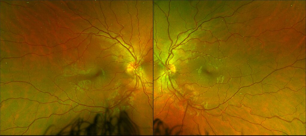
Optos
The Optos is a camera that can photograph the entire retina. This allows us to document and follow diseases such as retinal detachments and diabetic retinopathy. Really any retinal pathology can be documented with this.
Fluorescein Angiogram
The Optos can be used with vegetable dye injected into the vein to image, with great detail, the vascular system of the retina. This allows great ability to assess vascular pathology and precisely direct treatment. This is especially important with diabetic retinopathy and vascular occlusive disease as well as Macular degeneration.
Indocyanine Green (ICG)
This dye is used in conjunction with the Optos imaging system by injecting a different dye. This allows detections of diseases affecting the choroid, deeper than the retina. This is particularly helpful with inflammatory disorders of the posterior segment of the eye as well as Macular degeneration.
Overall, we are very fortunate in ophthalmology. We are able to visualize most of the anatomic areas of the eye allowing us better diagnosis, treatment and monitoring of treatment.